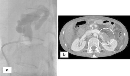

Grade 2–3 oesophageal varices were identified on oesophago-gastro-duodenoscopy, and computed tomography showed multiple right para-colic portosystemic collaterals around the hepatic flexure and ascending colon. Case presentationĪ 55-year-old man with background of alcoholic liver cirrhosis presented with per-rectal bleeding due to caecal varices. However, there are only two other cases that have reported successfully treating colonic varices using balloon-occluded retrograde transvenous obliteration (BRTO), an endovascular procedure typically performed for gastric varices. While established guidelines exist for the management of oesophageal and gastric variceal bleeding, this is currently lacking for colonic varices.īeta-blockers, transjugular intrahepatic portosystemic shunt insertion and subtotal colectomy have been reported as management methods.

They can be located in the duodenum, small intestines, colon or rectum, and may result in massive haemorrhage. Ectopic varices are uncommon and typically due to underlying liver cirrhosis.


 0 kommentar(er)
